File:Tetraspora gelatinosa.jpg

原始文件 (1,047 × 700像素,文件大小:139 KB,MIME类型:image/jpeg)
摘要
| 描述 |
Identifier: algvolimyxophy00west Title: Algæ. Vol. I. Myxophyceæ, Peridinieæ, Bacillarieæ, Chlorophyceæ, together with a brief summary of the occurrence and distribution of freshwat4er Algæ Year: 1916 (1910s) Authors: West, G. S. (George Stephen), 1876-1919 Subjects: Algae Publisher: Cambridge [Eng.] The University press Contributing Library: MBLWHOI Library Digitizing Sponsor: MBLWHOI Library
Click here to view book online to see this illustration in context in a browseable online version of this book.
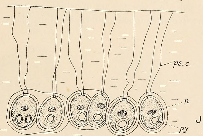
Text Appearing After Image: Fig. 113. A-G, Schizochlamys gelatinosa A. Br. A, vegetative cell showing pseudocilia, ×625; B, cell showing ecdysis of outer layers of wall, ×415; C and D, formation of zoogonidia, ×625; E, zoogonidium, ×830; F and G, zoogonidium changing to Schizochlamys-cell, ×830. H and I, Apiocystis brauniana Näg. H, pear-shaped colony, ×430; I, three cells showing pseudocilia, b, two daughter-cells from a division, the second pseudocilium not yet developed, ×860. J, Tetraspora gelatinosa (Vauch.) Desv., periphery of colony showing a few of the cells with their pseudocilia, × about 900. cv, contractile vacuole; n, nucleus; ol, oil globule; ps.c., pseudocilia; py, pyrenoid ; sf, stigma (or pigment-spot). (A-G, after Scherffel; J, after Chodat.) colony. The cells multiply by repeated division, chiefly in two directions inone plane, with the conversion of the walls of the mother-cells into mucilage.The pseudocilia are embedded in the mucilage of the colony (fig. 113 J), andeach cell is o
|
| 日期 | |
| 来源 | Image from page 200 of "Algæ. Vol. I. Myxophyceæ, Peridinieæ, Bacillarieæ, Chlorophyceæ, together with a brief summary of the occurrence and distribution of freshwat4er Algæ" (1916) |
| 作者 | Internet Archive Book Images |
| 授权 (二次使用本文件) |
Internet Archive Book Images @ Flickr Commons |
| 其他版本 |
.jpg)
许可协议
本图像是从Flickr的公众相簿计画取得而来。该上传组织可能有多种理由认定该影像无已知版权限制,例如:
更多资讯请参见 https://flickr.com/commons/usage/ 如果该图像拥有特定且决定性的版权资讯,请在该影像新增额外的版权标签。更多资讯请参见Commons:Licensing。 |
许可协议
|
本作品在其来源国以及其他著作权期限是作者逝世后100年或以下的国家和地区属于公有领域。 | |
| 本文件已被确认为免除已知的著作权法限制(包括所有相关权利)。 | |
文件历史
点击某个日期/时间查看对应时刻的文件。
| 日期/时间 | 缩略图 | 大小 | 用户 | 备注 | |
|---|---|---|---|---|---|
| 当前 | 2019年10月3日 (四) 14:06 | | 1,047 × 700(139 KB) | Awkwafaba | File:Image from page 200 of "Algæ. Vol. I. Myxophyceæ, Peridinieæ, Bacillarieæ, Chlorophyceæ, together with a brief summary of the occurrence and distribution of freshwat4er Algæ" (1916).jpg cropped 35 % horizontally, 57 % vertically using CropTool with lossless mode. |
.jpg)
文件用途
以下页面使用本文件:
全域文件用途
以下其他wiki使用此文件:
- ceb.wikipedia.org上的用途
- fr.wikipedia.org上的用途
- species.wikimedia.org上的用途
- tr.wikipedia.org上的用途
- www.wikidata.org上的用途