File:Epidermis-delimited.JPG

本预览的尺寸:800 × 496像素。 其他分辨率:320 × 198像素 | 640 × 397像素 | 1,000 × 620像素。
原始文件 (1,000 × 620像素,文件大小:240 KB,MIME类型:image/jpeg)
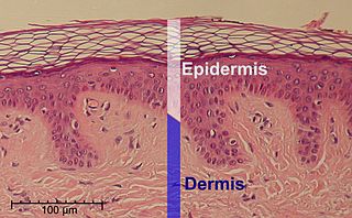
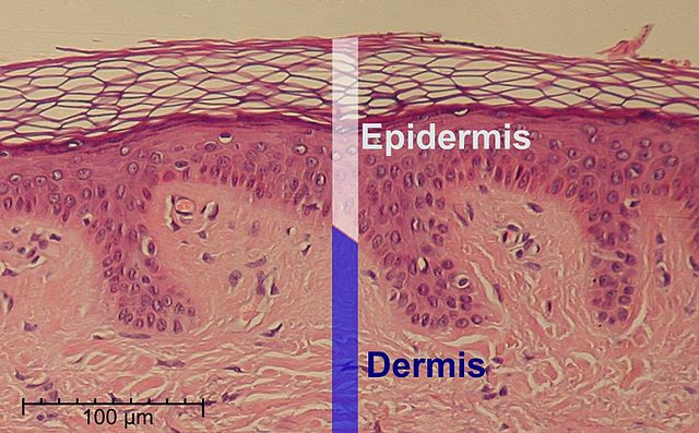
| 描述 | This is a hematoxylin and eosin stained slide at 10x of normal epidermis. | ||
| 日期 | |||
| 来源 |
Scale at lower left was created from the an estimation of mean epidermal cell nuclei of 8.6 μm according to the following study:
|
||
| 作者 | Cropped and labeled by Fama Clamosa (talk) and Mikael Häggström, respectively | ||
| 授权 (二次使用本文件) |
我,本作品著作权人,特此采用以下许可协议发表本作品:
|
| 这是一张修改过的图片,这意味着它已在原版本的基础上通过软件进行了编辑,改动内容:cropped。其原始版本为:Normal Epidermis and Dermis with Intradermal Nevus 10x.JPG。
|
文件历史
点击某个日期/时间查看对应时刻的文件。
| 日期/时间 | 缩略图 | 大小 | 用户 | 备注 | |
|---|---|---|---|---|---|
| 当前 | 2014年5月31日 (六) 16:48 | | 1,000 × 620(240 KB) | Mikael Häggström | Corrected scale |
| 2014年5月31日 (六) 16:29 |  | 1,000 × 620(240 KB) | Mikael Häggström | +Scale | |
| 2011年5月27日 (五) 16:18 |  | 1,000 × 620(238 KB) | Mikael Häggström | Improved | |
| 2010年6月29日 (二) 14:38 |  | 1,000 × 620(244 KB) | Mikael Häggström | {{Information |Description={{en|1=f}} |Source={{own}} |Author=Mikael Häggström |Date=f |Permission= |other_versions= }} |
文件用途
以下页面使用本文件:
全域文件用途
以下其他wiki使用此文件:
- af.wikipedia.org上的用途
- ar.wikipedia.org上的用途
- bn.wikipedia.org上的用途
- bs.wikipedia.org上的用途
- ca.wikipedia.org上的用途
- cv.wikipedia.org上的用途
- de.wikipedia.org上的用途
- el.wikipedia.org上的用途
- en.wikipedia.org上的用途
- en.wikibooks.org上的用途
- es.wikipedia.org上的用途
- fa.wikipedia.org上的用途
- fr.wikipedia.org上的用途
- gl.wikipedia.org上的用途
- gl.wiktionary.org上的用途
- ht.wikipedia.org上的用途
- hu.wikipedia.org上的用途
- it.wikibooks.org上的用途
- la.wikipedia.org上的用途
- myv.wikipedia.org上的用途
- pl.wikipedia.org上的用途
- pt.wikipedia.org上的用途
- ro.wikipedia.org上的用途
- ta.wikipedia.org上的用途
- te.wikibooks.org上的用途
- vi.wikipedia.org上的用途
- war.wikipedia.org上的用途
- www.wikidata.org上的用途