File:Mitochondria, mammalian lung - TEM.jpg
此为最大尺寸。
Mitochondria,_mammalian_lung_-_TEM.jpg (640 × 480像素,文件大小:96 KB,MIME类型:image/jpeg)
摘要
| 描述 |
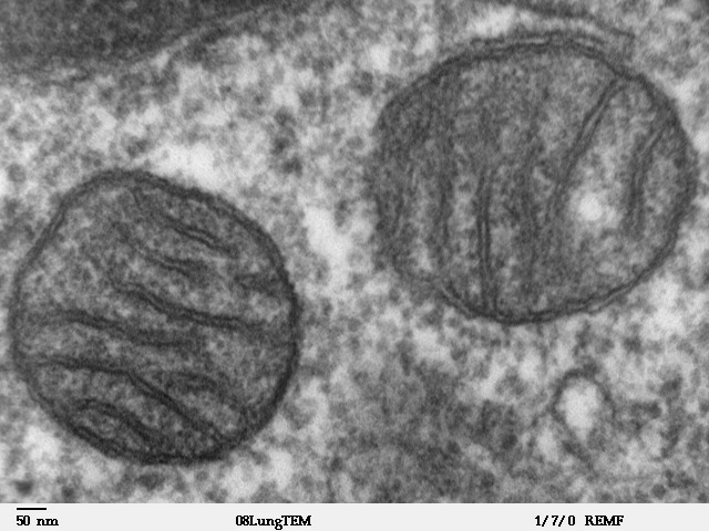
Transmission electron microscope image of a thin section cut through an area of mammalian lung tissue. The high magnification image shows two mitochondria. JEOL 100CX TEM |
| 来源 | |
| 作者 | Louisa Howard |
| 授权 (二次使用本文件) |
PD |
许可协议
| 本作品已被作者Louisa Howard释出到公有领域。这适用于全世界。 在一些国家这可能不合法;如果是这样的话,那么: Louisa Howard无条件地授予任何人以任何目的使用本作品的权利,除非这些条件是法律规定所必需的。
|
文件历史
点击某个日期/时间查看对应时刻的文件。
| 日期/时间 | 缩略图 | 大小 | 用户 | 备注 | |
|---|---|---|---|---|---|
| 当前 | 2008年5月16日 (五) 15:09 | | 640 × 480(96 KB) | Vojtěch Dostál | Reverted to version as of 15:37, 5 October 2006, my fault |
| 2008年5月16日 (五) 12:51 |  | 640 × 433(84 KB) | Vojtěch Dostál | {{Information |Description=Transmission electron microscope image of a thin section cut through an area of mammalian lung tissue. The high magnification image shows a mitochondria. JEOL 100CX TEM |Source= * http://remf.dartmouth.edu/imagesindex.html * h | |
| 2008年5月16日 (五) 12:47 |  | 640 × 453(86 KB) | Vojtěch Dostál | {{Information |Description=Transmission electron microscope image of a thin section cut through an area of mammalian lung tissue. The high magnification image shows a mitochondria. JEOL 100CX TEM |Source= * http://remf.dartmouth.edu/imagesindex.html * h | |
| 2006年10月5日 (四) 15:37 |  | 640 × 480(96 KB) | Patho | {{Information |Description=Transmission electron microscope image of a thin section cut through an area of mammalian lung tissue. The high magnification image shows a mitochondria. JEOL 100CX TEM |Source= * http://remf.dartmouth.edu/imagesindex.html * h |
文件用途
全域文件用途
以下其他wiki使用此文件:
- ar.wikipedia.org上的用途
- az.wiktionary.org上的用途
- be.wikipedia.org上的用途
- bg.wikipedia.org上的用途
- bn.wikipedia.org上的用途
- br.wikipedia.org上的用途
- bs.wikipedia.org上的用途
- ca.wikipedia.org上的用途
- cdo.wikipedia.org上的用途
- da.wikipedia.org上的用途
- de.wikibooks.org上的用途
- el.wikipedia.org上的用途
- en.wikipedia.org上的用途
- en.wikibooks.org上的用途
- en.wikiversity.org上的用途
- User:Jtwsaddress42/Projects/Project 1
- User:Jtwsaddress42/Projects/Project 1/Parts
- User:Jtwsaddress42/Projects/Project 1/Parts/Part 3
- User:Jtwsaddress42/Projects/Project 1/Chapters/Chapter 10
- User:Jtwsaddress42/Projects/Project 1/Sections/Chapter 10/Phase II - The Oxygen Crisis and the Rise of the Aerobic Bioshphere (1.9-0.95 bya)
- User:Jtwsaddress42/Clade
- User:Jtwsaddress42/Clade/Gracilicutes to Proteobacteria
- en.wiktionary.org上的用途
- es.wikipedia.org上的用途
- et.wikipedia.org上的用途
- eu.wikipedia.org上的用途
- ext.wikipedia.org上的用途
- fa.wikipedia.org上的用途
- fr.wikipedia.org上的用途
查看本文件的更多全域用途。