File:PET-image.jpg

本预览的尺寸:679 × 600像素。 其他分辨率:272 × 240像素 | 543 × 480像素 | 869 × 768像素 | 1,132 × 1,000像素。
原始文件 (1,132 × 1,000像素,文件大小:139 KB,MIME类型:image/jpeg)
摘要
| 描述 |
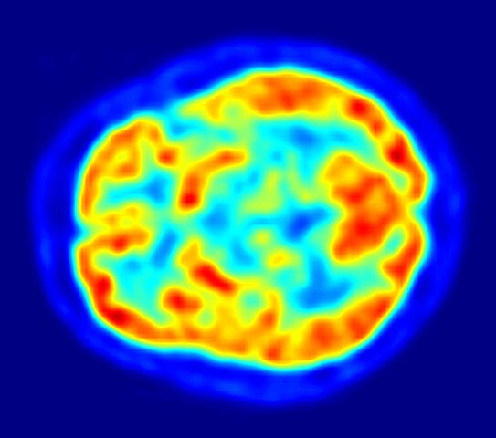
English: This is a transaxial slice of the brain of a 56 year old patient (male) taken with positron emission tomography (PET). The injected dose have been 282 MBq of 18F-FDG and the image was generated from a 20 minutes measurement with an ECAT Exact HR+ PET Scanner. Red areas show more accumulated tracer substance (18F-FDG) and blue areas are regions where low to no tracer have been accumulated.
العربية: صورة مقطعية للدماغ البشري تظهر استهلاك الطاقة. |
||
| 日期 | |||
| 来源 | 自己的作品 | ||
| 作者 | Jens Maus (http://jens-maus.de/) | ||
| 授权 (二次使用本文件) |
|
文件历史
点击某个日期/时间查看对应时刻的文件。
| 日期/时间 | 缩略图 | 大小 | 用户 | 备注 | |
|---|---|---|---|---|---|
| 当前 | 2017年12月12日 (二) 02:00 | | 1,132 × 1,000(139 KB) | SteinsplitterBot | Bot: Image rotated by 270° |
| 2010年3月16日 (二) 14:36 |  | 1,002 × 1,132(134 KB) | Damato | uploaded another PET image with a higher resolution which might be more usable for printing it and which has a better color scale. | |
| 2005年11月7日 (一) 09:47 |  | 373 × 405(48 KB) | Damato | This is an image taken from a typical PET acquisition. It is a tomographic view of a brain examination in transaxial view. Red areas show more accumulated radioactivity and blue areas are partions where low to no activity was accumulated. It should illust |
文件用途
全域文件用途
以下其他wiki使用此文件:
- ar.wikipedia.org上的用途
- arz.wikipedia.org上的用途
- ast.wikipedia.org上的用途
- bg.wikipedia.org上的用途
- bn.wikipedia.org上的用途
- ca.wikipedia.org上的用途
- de.wikipedia.org上的用途
- de.wikibooks.org上的用途
- el.wikipedia.org上的用途
- en.wikipedia.org上的用途
- Positron emission tomography
- Neurolinguistics
- Human brain
- Scintigraphy
- Timeline of tuberous sclerosis
- User:Portakalsinatra
- Wikipedia:Wikipedia Signpost/2011-03-07/Features and admins
- User talk:Silver seren/Archive 10
- Childhood acquired brain injury
- User:Rkasinadhuni3/practice sandbox
- User:Mcorrin3/Sandbox Practice
- User:LoriJeanMarie/Brain science practice page
- User:Gilyardterence/Pediatric Acquired Brain Injury
- Wikipedia:Wikipedia Signpost/Single/2011-03-07
- Wikipedia:WikiProject Cannabis/Members
- User:Anthonyhcole/Parkinson's disease
- User:Silver seren/Barnstars
- User:Flyer22 Frozen/Human brain
- User:Cglife.bmarcus/WikiProjectCards/WikiProject Cannabis
- en.wikiquote.org上的用途
- en.wikiversity.org上的用途
- es.wikipedia.org上的用途
查看本文件的更多全域用途。