File:PET-schema.png

本预览的尺寸:800 × 586像素。 其他分辨率:320 × 235像素 | 640 × 469像素 | 1,024 × 750像素 | 1,280 × 938像素。
原始文件 (1,280 × 938像素,文件大小:698 KB,MIME类型:image/png)
摘要
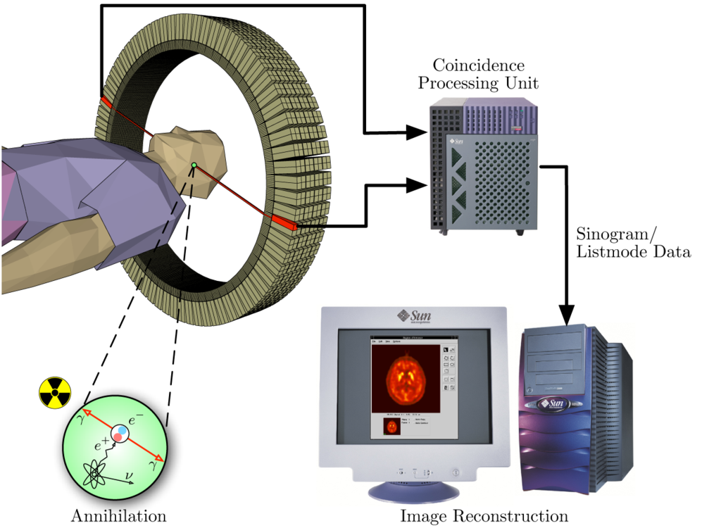
| 描述 | The image illustrates the processing principles of a positron emission tomograph (PET) commonly used in cancer diagnostics. It shows how during the annihilation process two photons are emitted in diametrically opposite directions. These photons are registered by the scanner as soon as they arrive at the detector ring. After the registration, the data is forwarded to a processing unit which decides if two registered events are selected as a so-called coincidence event. All coincidences are forwarded to the image processing unit where the final image data is produced via mathematical image reconstruction procedures. | ||
| 日期 | |||
| 来源 |
own work - part of master thesis |
||
| 作者 | Jens Maus (http://jens-maus.de/) | ||
| 授权 (二次使用本文件) |
|
文件历史
点击某个日期/时间查看对应时刻的文件。
| 日期/时间 | 缩略图 | 大小 | 用户 | 备注 | |
|---|---|---|---|---|---|
| 当前 | 2005年11月17日 (四) 15:25 | | 1,280 × 938(698 KB) | Damato | Uploaded a version with a higher resolution. Content not changed. |
| 2005年11月5日 (六) 08:34 |  | 449 × 329(122 KB) | Damato | Data processing feature of an tomographic acquisition using a positron emission tomograph. It illustrates the principials of a PET acquisition right from the annihilation process, through how the lines of response LOR are registered by the tom |
文件用途
以下页面使用本文件:
全域文件用途
以下其他wiki使用此文件:
- de.wikipedia.org上的用途
- en.wikipedia.org上的用途
- eo.wikipedia.org上的用途
- es.wikipedia.org上的用途
- eu.wikipedia.org上的用途
- fa.wikipedia.org上的用途
- fr.wikipedia.org上的用途
- fr.wikibooks.org上的用途
- fr.wikiversity.org上的用途
- he.wikipedia.org上的用途
- hi.wikipedia.org上的用途
- hu.wikipedia.org上的用途
- is.wikipedia.org上的用途
- it.wikipedia.org上的用途
- kn.wikipedia.org上的用途
- nl.wikipedia.org上的用途
- pl.wikipedia.org上的用途
- pt.wikipedia.org上的用途
- ro.wikipedia.org上的用途
- sr.wikipedia.org上的用途
- th.wikipedia.org上的用途
- uz.wikipedia.org上的用途