File:Staphylococcus aureus VISA 2.jpg

原始文件 (1,420 × 1,091像素,文件大小:259 KB,MIME类型:image/jpeg)
| 描述 |
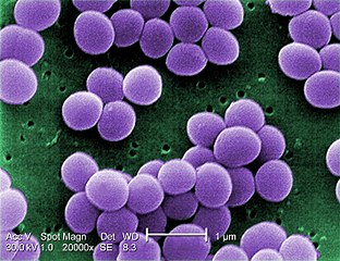
English: Under a very high magnification of 20,000x, this scanning electron micrograph (SEM) shows a strain of Staphylococcus aureus bacteria taken from a vancomycin intermediate resistant culture (VISA). Under SEM, one can not tell the difference between bacteria that are susceptible, or multidrug resistant, but with transmission electron microscopy (TEM), VISA isolates exhibit a thickening in the cell wall that may attribute to their reduced susceptibility to vancomycin . See PHIL 11156 for a black and white version of this image. VISA and VRSA are specific types of antimicrobial-resistant staph bacteria. While most staph bacteria are susceptible to the antimicrobial agent vancomycin some have developed resistance. VISA and VRSA cannot be successfully treated with vancomycin because these organisms are no longer susceptibile to vancomycin. However, to date, all VISA and VRSA isolates have been susceptible to other Food and Drug Administration (FDA) approved drugs. How do VISA and VRSA get their names? Staph bacteria are classified as VISA or VRSA based on laboratory tests. Laboratories perform tests to determine if staph bacteria are resistant to antimicrobial agents that might be used for treatment of infections. For vancomycin and other antimicrobial agents, laboratories determine how much of the agent it requires to inhibit the growth of the organism in a test tube. The result of the test is usually expressed as a minimum inhibitory concentration (MIC) or the minimum amount of antimicrobial agent that inhibits bacterial growth in the test tube. Therefore, staph bacteria are classified as VISA if the MIC for vancomycin is 4-8µg/ml, and classified as VRSA if the vancomycin MIC is >16µg/ml. |
||
| 日期 | |||
| 来源 |
|
||
| 作者 |
Content Providers(s): CDC/ Matthew J. Arduino, DRPH |
||
| 授权 (二次使用本文件) |
PD-USGov-HHS-CDC English: None - This image is in the public domain and thus free of any copyright restrictions. As a matter of courtesy we request that the content provider be credited and notified in any public or private usage of this image. |
|
|
文件历史
点击某个日期/时间查看对应时刻的文件。
| 日期/时间 | 缩略图 | 大小 | 用户 | 备注 | |
|---|---|---|---|---|---|
| 当前 | 2009年8月4日 (二) 02:24 | | 1,420 × 1,091(259 KB) | Raeky | {{Information |Description={{en|1='''Under a very high magnification of 20,000x, this scanning electron micrograph (SEM) shows a strain of Staphylococcus aureus bacteria taken from a vancomycin intermediate resistant culture (VISA).'''<p> Under SEM, one |
文件用途
以下48个页面使用本文件:
- F质粒
- Γ-變形菌
- 云南链霉菌
- 伯氏疏螺旋體
- 保加利亚乳杆菌
- 分歧焦蟲
- 厭氧氨氧化菌
- 发酵厌氧杆菌属
- 嗜碱海洋菌属
- 實驗室黴漿菌
- 居鼻鲍登氏菌
- 弗兰克氏菌科
- 性菌毛
- 抗輻射奇異球菌
- 擬態弧菌
- 新月柄杆菌
- 枯草桿菌
- 海南链霉菌
- 潮滩食琼脂菌
- 熱帶魚弧菌
- 熱微菌門
- 熱脫硫桿菌門
- 異常球菌-棲熱菌門
- 白色食琼脂菌
- 磁小体
- 立氏立克次體
- 紫細菌
- 細弱螺旋體
- 結核桿菌
- 網團菌門
- 聚球藻菌属
- 芽孢桿菌綱
- 荚膜
- 螺旋體門
- 豬鏈球菌
- 路鄧葡萄球菌
- 酸微菌亞綱
- 酸桿菌目
- 金黃色葡萄球菌
- 霍亂弧菌
- 類鼻疽伯克氏菌
- 食琼脂科维尔氏菌
- 食琼脂链状小卵菌
- 香港鷗桿菌
- 鼻疽伯克霍爾德氏菌
- User:Kenchan hihi
- User:Whw219
- Template:Bacteria-stub
全域文件用途
以下其他wiki使用此文件:
- ar.wikipedia.org上的用途
- فيلقية
- أشعار بكتيرية
- آزوتية
- ريكتسيا
- مستجذرة
- مفطورة
- ملوية (جنس)
- وتدية خناقية
- متفطرة
- مطثية حاطمة
- هدب (بكتيريا)
- شعاوات
- متفطرة جذامية
- مرق السيلينيت
- بوريليا برغدورفيرية
- مكورات
- صبغة أورامين رومدامين
- اختبار الأكسيداز
- انزلاق بكتيري
- ملتويات (بكتيريا)
- ملتوية معوية
- نستق
- قالب:بذرة بكتيريا
- نوستك
- مستدمية
- كليبسيلا
- سفينغوبيوم
- جرثومة مخاطية
- متقلبات
- متقلبات زيتا
- متقلبات إيبسيلونية
- متقلبات غاما
- متقلبات بيتا
- متقلبات ألفا
- بكتيريا مغزلية
- عصوانيات
- خضربيات
- دليل برجاي لعلم الجراثيم المنهجي
- جراثيم ثؤلولية
- مستعلقات
- سحناوات
- متمصرة
- بكتيريا حمضية
- متدثرات
- كلورو بكتيريا
- شبكيات الكبب
- شبكية الكبة حرارية
- ليفيات
- سلسلية
- سلسلية طوقية الشكل
查看本文件的更多全域用途。