File:Virus Replication.svg

原始文件 (SVG文件,尺寸为462 × 426像素,文件大小:205 KB)
摘要
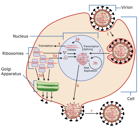
| 描述 | A diagram of influenza viral cell invasion and replication. |
| 日期 | |
| 来源 | Redrawn from w:Image:Virusreplication.png using Adobe Illustrator. |
| 作者 | User:YK Times |
| 其他版本 |
 |


|
此SVG檔案包含可翻譯至您的語言的内嵌文字,可以使用任何可用SVG編輯器、文本編輯器或SVG翻譯工具來翻譯。詳情請見:關於翻譯SVG檔案。 |
Description from Scheme of Influenza A virus replication (NCBI): "A virion attaches to the host cell membrane via HA and enters the cytoplasm by receptor-mediated endocytosis (STEP 1), thereby forming an endosome. A cellular trypsin-like enzyme cleaves HA into products HA1 and HA2 (not shown). HA2 promotes fusion of the virus envelope and the endosome membranes. A minor virus envelope protein M2 acts as a ion channel thereby making the inside of the virion more acidic. As a result, the major envelope protein M1 dissociates from the nucleocapsid and vRNPs are translocated into the nucleus (STEP 2) via interaction between NP and cellular transport machinery. In the nucleus, the viral polymerase complexes transcribe (STEP 3a) and replicate (STEP 3b) the vRNAs. Newly synthesized mRNAs migrate to cytoplasm (STEP 4) where they are translated. Posttranslational processing of HA, NA, and M2 includes transportation via Golgi apparatus to the cell membrane (STEP 5b). NP, M1, NS1 (nonstructural regulatory protein - not shown) and NEP (nuclear export protein, a minor virion component - not shown) move to the nucleus (STEP 5a) where they bind freshly synthesized copies of vRNAs. The newly formed nucleocapsids migrate into the cytoplasm in a NEP-dependent process and eventually interact via M1 with a region of the cell membrane where HA, NA and M2 have been inserted (STEP 6). Then the newly synthesized virions bud from infected cell (STEP 7). NA destroys the sialic acid moiety of cellular receptors, thereby releasing the progeny virions."
许可协议

|
已授权您依据自由软件基金会发行的无固定段落及封面封底文字(Invariant Sections, Front-Cover Texts, and Back-Cover Texts)的GNU自由文件许可协议1.2版或任意后续版本的条款,复制、传播和/或修改本文件。该协议的副本请见“GNU Free Documentation License”。 |
| 本文件采用知识共享署名-相同方式共享 3.0 未本地化版本许可协议授权。 | ||
| ||
| 本许可协议标签作为GFDL许可协议更新的组成部分被添加至本文件。 |
- 您可以自由地:
- 共享 – 复制、发行并传播本作品
- 修改 – 改编作品
- 惟须遵守下列条件:
- 署名 – 您必须对作品进行署名,提供授权条款的链接,并说明是否对原始内容进行了更改。您可以用任何合理的方式来署名,但不得以任何方式表明许可人认可您或您的使用。
- 相同方式共享 – 如果您再混合、转换或者基于本作品进行创作,您必须以与原先许可协议相同或相兼容的许可协议分发您贡献的作品。
| 註解 | 該圖片含有註解:在維基媒體共享資源上查看註解 |
文件历史
点击某个日期/时间查看对应时刻的文件。
| 日期/时间 | 缩略图 | 大小 | 用户 | 备注 | |
|---|---|---|---|---|---|
| 当前 | 2007年3月6日 (二) 02:54 | | 462 × 426(205 KB) | YK Times | {{Information |Description=A diagram of influenza viral cell invasion and replication. |Source=Redrawn from w:Image:Virusreplication.png using Adobe Illustrator. |Date=March 5, 2007 |Author= User:YK Times |Permission= |other_versions=[[:w:Image:V |
文件用途
以下页面使用本文件:
全域文件用途
以下其他wiki使用此文件:
- bg.wikipedia.org上的用途
- bn.wikipedia.org上的用途
- br.wikipedia.org上的用途
- ca.wikipedia.org上的用途
- cs.wikipedia.org上的用途
- da.wikipedia.org上的用途
- de.wikipedia.org上的用途
- el.wikipedia.org上的用途
- en.wikipedia.org上的用途
- Orthomyxoviridae
- Amantadine
- Viral replication
- Viral life cycle
- Viral entry
- File:Virusreplication.png
- User:YK Times/Graphic Lab/examples
- Wikipedia:Graphics Lab/Images to improve/Archive/Mar 2007
- Viral shedding
- Template:Influenza virus life cycle
- Viral neuraminidase
- Tilapia tilapinevirus
- User:Zoe.gum/sandbox
- Wikipedia talk:WikiProject Viruses/Archive 4
- User:Anicm1/sandbox
- en.wikibooks.org上的用途
- es.wikipedia.org上的用途
- fa.wikipedia.org上的用途
- fr.wikipedia.org上的用途
- hy.wikipedia.org上的用途
- id.wikipedia.org上的用途
- it.wikipedia.org上的用途
- ja.wikipedia.org上的用途
- kk.wikipedia.org上的用途
- ko.wikipedia.org上的用途
查看本文件的更多全域用途。