File:Blood clot in scanning electron microscopy.jpg
此为最大尺寸。
Blood_clot_in_scanning_electron_microscopy.jpg (700 × 475像素,文件大小:76 KB,MIME类型:image/jpeg)
摘要
| 描述 |
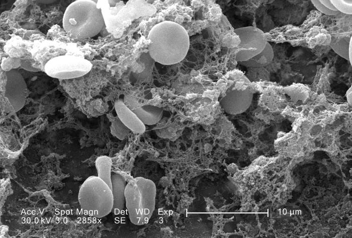
English: This scanning electron micrograph (SEM) depicted a number of red blood cells found enmeshed in a fibrinous matrix on the luminal surface of an indwelling vascular catheter; Magnified 2858x.
Note the biconcave cytomorphologic shape of each erythrocyte, which increases the surface area of these hemoglobin-filled cells, thereby, promoting a greater degree of gas exchange, which is their primary function in an in vivo setting. In their adult phase, these cells possess no nucleus. What appears to be irregularly-shaped chunks of debris, are actually fibrin clumps, which when inside the living organism, functions as a key component in the process of blood clot formation, acting to entrap the red blood cells in a mesh-like latticework of proteinaceous strands, thereby, stabilizing and strengthening the clot, in much the same way as rebar acts to strengthen, and reinforce cement. |
| 日期 | |
| 来源 | http://phil.cdc.gov/phil/details.asp |
| 作者 | Janice Carr |
许可协议
| 本作品已被作者http://phil.cdc.gov/phil/details.asp Janice Carr释出到公有领域。这适用于全世界。 在一些国家这可能不合法;如果是这样的话,那么: http://phil.cdc.gov/phil/details.asp Janice Carr无条件地授予任何人以任何目的使用本作品的权利,除非这些条件是法律规定所必需的。
|
文件历史
点击某个日期/时间查看对应时刻的文件。
| 日期/时间 | 缩略图 | 大小 | 用户 | 备注 | |
|---|---|---|---|---|---|
| 当前 | 2015年5月13日 (三) 17:25 | | 700 × 475(76 KB) | Jean-madeleine de sainte agathe | User created page with UploadWizard |
文件用途
以下页面使用本文件:
全域文件用途
以下其他wiki使用此文件:
- fr.wikipedia.org上的用途
- zh-yue.wikipedia.org上的用途