File:Bronchiolar epithelium 3 - SEM.jpg

本预览的尺寸:585 × 599像素。 其他分辨率:234 × 240像素 | 469 × 480像素 | 750 × 768像素 | 1,024 × 1,049像素。
原始文件 (1,024 × 1,049像素,文件大小:375 KB,MIME类型:image/jpeg)
摘要
| 描述 |
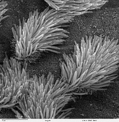
Scanning electron microscope image of lung trachea epithelium. There are both ciliated and non-ciliated cells in this epithelium. Note the difference in size between the cilia and the microvilli (on the non-ciliated cell surface). Zeiss DSM 962 SEM |
| 来源 | |
| 作者 | Charles Daghlian |
| 授权 (二次使用本文件) |
PD |
许可协议
| 本作品已被作者Charles Daghlian释出到公有领域。这适用于全世界。 在一些国家这可能不合法;如果是这样的话,那么: Charles Daghlian无条件地授予任何人以任何目的使用本作品的权利,除非这些条件是法律规定所必需的。
|
文件历史
点击某个日期/时间查看对应时刻的文件。
| 日期/时间 | 缩略图 | 大小 | 用户 | 备注 | |
|---|---|---|---|---|---|
| 当前 | 2006年10月7日 (六) 14:16 | | 1,024 × 1,049(375 KB) | Patho | {{Information |Description=Scanning electron microscope image of lung trachea epithelium. There are both ciliated and on-ciliated cells in this epithelium. Note the difference in size between the cilia and the microvilli(on non-ciliated cell surface) Zei |
文件用途
以下页面使用本文件:
全域文件用途
以下其他wiki使用此文件:
- ar.wikipedia.org上的用途
- ast.wikipedia.org上的用途
- bs.wikipedia.org上的用途
- ca.wikipedia.org上的用途
- cs.wikipedia.org上的用途
- da.wikipedia.org上的用途
- de.wikipedia.org上的用途
- de.wikibooks.org上的用途
- en.wikipedia.org上的用途
- es.wikipedia.org上的用途
- eu.wikipedia.org上的用途
- fa.wikipedia.org上的用途
- fr.wikipedia.org上的用途
- gl.wikipedia.org上的用途
- he.wikipedia.org上的用途
- he.wiktionary.org上的用途
- hi.wikipedia.org上的用途
- id.wikipedia.org上的用途
- jv.wikipedia.org上的用途
- kk.wikipedia.org上的用途
- lt.wikipedia.org上的用途
- lv.wikipedia.org上的用途
- ms.wikipedia.org上的用途
- nl.wikipedia.org上的用途
- nn.wikipedia.org上的用途
- no.wikipedia.org上的用途
- pl.wikipedia.org上的用途
- pl.wiktionary.org上的用途
- pt.wikipedia.org上的用途
- ru.wikipedia.org上的用途
- ru.wiktionary.org上的用途
- sh.wikipedia.org上的用途
查看本文件的更多全域用途。