File:Calcarine sulcus animation small.gif

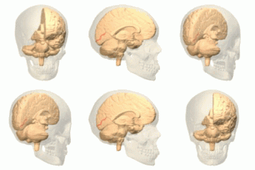
Calcarine_sulcus_animation_small.gif (300 × 300像素,文件大小:1.68 MB,MIME类型:image/gif、​循环、​72帧、​11秒)


描述
English: Calcarine sulcus of left hemisphere. One of sulcus located in medial wall. Polygon data are from BodyParts3D maintained by Database Center for Life Science(DBCLS).
日本語: 左大脳半球の鳥距溝。内側面に位置する脳溝のひとつ。 ポリゴンデータはDatabase Center for Life Science(DBCLS)の作成しているBodyParts3Dより。
日期
来源 Polygon data are from BodyParts3D[1]
作者 Polygon data were generated by Database Center for Life Science(DBCLS)[2].
授权
(二次使用本文件)
CC-BY-SA-2.1-jp
 
本图片使用Blender创作.
w:zh:知识共享
署名 相同方式共享
本文件采用知识共享署名-相同方式共享 2.1 日本许可协议授权。
您可以自由地:
  • 共享 – 复制、发行并传播本作品
  • 修改 – 改编作品
惟须遵守下列条件:
  • 署名 – 您必须对作品进行署名,提供授权条款的链接,并说明是否对原始内容进行了更改。您可以用任何合理的方式来署名,但不得以任何方式表明许可人认可您或您的使用。
  • 相同方式共享 – 如果您再混合、转换或者基于本作品进行创作,您必须以与原先许可协议相同或相兼容的许可协议分发您贡献的作品。
Anatomography
Anatomography
Database Center for Life Science (DBCLS)
This image was made out of, or made from, content published in a BodyParts3D/Anatomography web site. The content of their website is published under the Creative Commons Attribution 2.1 Japan license. The author and licenser of the contents is

You can download 3D-polygon data of whole human body. And you can also manipulate and edit the polygon data using 3D softwares, for example, Meshlab or Blender.

说明

添加一行文字以描述该文件所表现的内容

此文件中描述的项目

描繪內容

6123f4d03ae9a5d2bab824245283027ec285b3ee

1,759,684 字节

11.09999999999999

300 像素

300 像素

文件历史

点击某个日期/时间查看对应时刻的文件。

日期/时间缩⁠略⁠图大小用户备注
当前2012年9月18日 (二) 02:272012年9月18日 (二) 02:27版本的缩略图300 × 300(1.68 MB)Was a beemake it clearer
2010年1月11日 (一) 18:002010年1月11日 (一) 18:00版本的缩略图150 × 150(474 KB)Was a bee{{Information |Description={{en|1= Calcarine sulcus of left hemisphere. One of sulcus located in medial wall. Polygon data are from BodyParts3D maintained by Database Center for Life Science(DBCLS). }} {{ja|1=左大脳半球の鳥距溝。内側面に位�

以下页面使用本文件:

全域文件用途

以下其他wiki使用此文件: