File:Electron micrograph of neuromuscular junction (cross-section).jpg
此为最大尺寸。
Electron_micrograph_of_neuromuscular_junction_(cross-section).jpg (433 × 289像素,文件大小:95 KB,MIME类型:image/jpeg)
.jpg?uselang=zh)
.jpg?uselang=zh)
.jpg?action=history&uselang=zh)
.jpg)
摘要
| 描述 |
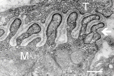
English: Electron micrograph showing a cross-section through the neuromuscular junction. T is the axon terminal, M is the muscle fiber. The arrow shows junctional folds with basal lamina. Postsynaptic densities are visible on the tips between the folds. The scale is 0.3 µm. |
| 日期 | Originally uploaded to en.wikipedia on 2006年3月10日. |
| 来源 | Synapse Web at the National Institute of Mental Health, National Institutes of Health; originally from en.wikipedia; description page is/was here. |
| 作者 | National Institute of Mental Health; originally uploaded by Nrets at en.wikipedia. |
许可协议
這個圖像是美國衛生與公眾服務部下屬的美國國立衛生研究院的作品。作為美國聯邦政府的作品,這個圖像屬於公有領域。
 |
||
| 本文件已被确认为免除已知的著作权法限制(包括所有相关权利)。 | ||
原始上传日志
(All user names refer to en.wikipedia)
- 2006-03-10 20:07 Nrets 433×289×8 (97758 bytes) Electron micrograph showing a cross section through the neuromuscular junction. T is the axon terminal, M is the muscle fiber. The arrow shows junctional folds with basal lamina. Postsynaptic densities are visible on the tips between the folds. Scale is 0
文件历史
点击某个日期/时间查看对应时刻的文件。
| 日期/时间 | 缩略图 | 大小 | 用户 | 备注 | |
|---|---|---|---|---|---|
| 当前 | 2007年3月22日 (四) 03:41 | | 433 × 289(95 KB) | Fran Rogers | {{Information |Description=Electron micrograph showing a cross section through the neuromuscular junction. T is the axon terminal, M is the muscle fiber. The arrow shows junctional folds with basal lamina. Postsynaptic densities are visible on the tips be |
文件用途
以下页面使用本文件:
全域文件用途
以下其他wiki使用此文件:
- ar.wikipedia.org上的用途
- cs.wikipedia.org上的用途
- de.wikipedia.org上的用途
- en.wikipedia.org上的用途
- es.wikipedia.org上的用途
- fa.wikipedia.org上的用途
- gl.wikipedia.org上的用途
- he.wikipedia.org上的用途
- ko.wikipedia.org上的用途
- pt.wikipedia.org上的用途
- ru.wikipedia.org上的用途
- uk.wikipedia.org上的用途
.jpg)