原始文件(805 × 949像素,文件大小:278 KB,MIME类型:image/gif


摘要

描述
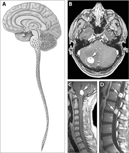
English: Distribution of Hemangioblastomas in the Central Nervous Systems of Study Patients

(A) Schematic representation of the distribution of CNS hemangioblastomas (red dots) in the 25 von Hippel-Lindau disease patients on MRI. Most (98%) of hemangioblastomas were found below the level of the tentorium in the cerebellum, brainstem, and spinal cord.

(B–D) Contrast-enhanced MRI demonstrating representative locations of hemangioblastomas including the cerebellum (B), brainstem (C) and spinal cord (D). (B) Axial view through the cerebellum demonstrating a hyperintense enhancing hemangioblastoma (arrow) with surrounding edema (hypointense area surrounding the tumor) that frequently is associated with these lesions. (C) Sagittal view through the posterior fossa demonstrating a hyperintense enhancing brainstem (medullary) hemangioblastoma (arrow) with surrounding edema. (D) Sagittal view through the thoracic and lumbar spinal cord demonstrating two hyperintense enhancing hemangioblastomas (arrows). The superior tumor is associated with a large intraspinal cyst (syrinx) that is common with these neoplasms (arrowhead)
来源 http://medicine.plosjournals.org/perlserv/?request=get-document&doi=10.1371/journal.pmed.0040060
作者

许可协议

w:zh:知识共享
署名
本文件采用知识共享署名 2.5 通用许可协议授权。
您可以自由地:
  • 共享 – 复制、发行并传播本作品
  • 修改 – 改编作品
惟须遵守下列条件:
  • 署名 – 您必须对作品进行署名,提供授权条款的链接,并说明是否对原始内容进行了更改。您可以用任何合理的方式来署名,但不得以任何方式表明许可人认可您或您的使用。
PLOS
PLOS
此文件发布于《公共科学图书馆》学术期刊中,其网站除特别指出外,其旗下的所有文章均以知识共享署名 4.0 许可协议(或发布时指定的版本)发布。

说明

添加一行文字以描述该文件所表现的内容

此文件中描述的项目

描繪內容

文件历史

点击某个日期/时间查看对应时刻的文件。

日期/时间缩⁠略⁠图大小用户备注
当前2007年5月31日 (四) 13:432007年5月31日 (四) 13:43版本的缩略图805 × 949(278 KB)Filip emDistribution of Hemangioblastomas in the Central Nervous Systems of Study Patients (A) Schematic representation of the distribution of CNS hemangioblastomas (red dots) in the 25 von Hippel-Lindau disease patients on MRI. Most (98%) of hemangioblastomas w

以下2个页面使用本文件:

全域文件用途

以下其他wiki使用此文件: