File:Wernicke's area animation.gif
此为最大尺寸。
Wernicke's_area_animation.gif (300 × 300像素,文件大小:1.99 MB,MIME类型:image/gif、循环、72帧、10秒)
| 描述 |
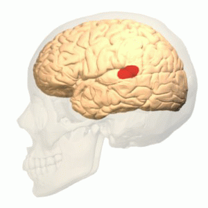
English: Wernicke's area (shown in red).
Colored region is posterior section of the superior temporal gyrus (pSTG) of the left cerebral hemisphere. Though this region is generally treated as Wernicke's area, there are many researches and discussions about its exact size and anatomical boundaries.
日本語: ウェルニッケ野。赤色で示す。 赤色の領域は左大脳半球の上側頭回の後部。現在一般にこの領域がウェルニッケ野として扱われているが、ウェルニッケ野の広さとその解剖学的境界については、現在も多くの研究と議論が行われている。
|
| 日期 | |
| 来源 | Polygon data are from BodyParts3D[1] |
| 作者 | Polygon data were generated by Database Center for Life Science(DBCLS)[2]. |
| 授权 (二次使用本文件) |
CC-BY-SA-2.1-jp |
本图片使用Blender创作.
本文件采用知识共享署名-相同方式共享 2.1 日本许可协议授权。
 |
 This image was made out of, or made from, content published in a BodyParts3D/Anatomography web site. The content of their website is published under the Creative Commons Attribution 2.1 Japan license. The author and licenser of the contents is
You can download 3D-polygon data of whole human body. And you can also manipulate and edit the polygon data using 3D softwares, for example, Meshlab or Blender. |
文件历史
点击某个日期/时间查看对应时刻的文件。
| 日期/时间 | 缩略图 | 大小 | 用户 | 备注 | |
|---|---|---|---|---|---|
| 当前 | 2014年5月3日 (六) 09:36 | | 300 × 300(1.99 MB) | Was a bee | {{Information |Description={{en|1= Wernicke's area (shown in red).<br />Colored region is posterior section of the superior temporal gyrus (pSTG) of the left cerebral hemisphere. Though this region is generally treated as Wernic... |
文件用途
以下页面使用本文件:
全域文件用途
以下其他wiki使用此文件:
- en.wikipedia.org上的用途
- es.wikipedia.org上的用途
- fa.wikipedia.org上的用途
- fr.wikipedia.org上的用途
- hy.wikipedia.org上的用途
- it.wikipedia.org上的用途
- ja.wikipedia.org上的用途
- pt.wikipedia.org上的用途
- sv.wikipedia.org上的用途
- tr.wikipedia.org上的用途
- zh-yue.wikipedia.org上的用途