File:FIPCytology2.jpg
此為最大尺寸。
FIPCytology2.jpg (350 × 234 像素,檔案大小:40 KB,MIME 類型:image/jpeg)
摘要
| 描述 |
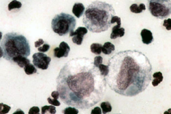
English: Color micrograph of the cytology of FIP-induced effusion. Magnification not specified; estimated to be 1000x.
Original caption: "The cytology of FIP effusion usually contains neutrophils, macrophages and lymphocytes." Image from "Feline Infectious Peritonitis: An Overview of Disease Transmission, Pathogenesis, Signs and Treatment With Emphasis on Diagnosis" ([1]) Clinical Pathology Clerkship Program |
| 日期 | 2005年九月30日 (原始上傳日期) |
| 來源 | 本檔案是從en.wikipedia轉移到維基共享資源。 |
| 作者 | 原上傳者為英文維基百科的Bk0 |
授權條款

|
本檔案的著作權持有者,在註明所有人姓名的前提下,允許任何人使用本檔案於任何用途。包含再散佈、衍生作品、商業用途及其他用途。 |
|
|
原始上傳日誌
原始描述頁面位於這裡。下列使用者名稱均來自en.wikipedia。
- 2005-09-30 00:14 Bk0 350×234×8 (40687 bytes) Color micrograph of the cytology of [[Feline infectious peritonitis|FIP]]-induced effusion. Magnification not specified; estimated to be 1000x. Original caption: "The cytology of FIP effusion usually contains neutrophils, macrophages and lymphocytes." I
檔案歷史
點選日期/時間以檢視該時間的檔案版本。
| 日期/時間 | 縮圖 | 尺寸 | 用戶 | 備註 | |
|---|---|---|---|---|---|
| 目前 | 2007年12月29日 (六) 18:53 | | 350 × 234(40 KB) | Euthygenes | {{Information |Description={{en|Color micrograph of the cytology of FIP-induced effusion. Magnification not specified; estimated to be 1000x. Original caption: "The cytology of FIP effusion usually contains neutrophi |
檔案用途
下列頁面有用到此檔案:
全域檔案使用狀況
以下其他 wiki 使用了這個檔案:
- el.wikipedia.org 的使用狀況
- en.wikipedia.org 的使用狀況
- et.wikipedia.org 的使用狀況
- fr.wikipedia.org 的使用狀況
- hu.wikipedia.org 的使用狀況
- ko.wikipedia.org 的使用狀況
- tr.wikipedia.org 的使用狀況