File:Cajal Retina.jpg
原始文件 (500 × 745像素,文件大小:83 KB,MIME类型:image/jpeg)
摘要
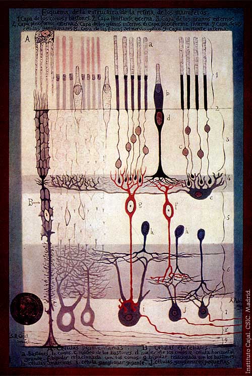
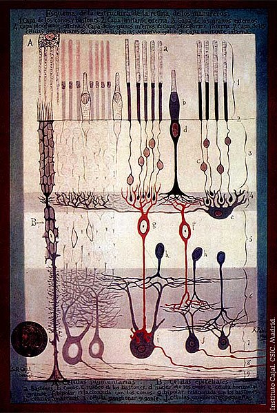
From "Structure of the Mammalian Retina" c.1900 By Santiago Ramon y Cajal.
Outline of the structure of the mammalian retina. 1. Rod and cone layer. 2. External limiting membrane. 3. Outer granular layer. 4. Outer plexiform layer. 5. Inner granular layer. 6. Inner plexiform layer. 7. Ganglion cell layer. 8. Optic nerve fibre layer. 9. Internal limiting membrane. A. Pigmented cells. B. Epithelial cells. a. Rods. b. Cones. c. Rod nucleus. d. Cone Nucleus. e. Large horizontal cell f. Cone-associated bipolar cell. g. Rod-associated bipolar cell. h. Amacrine cells. i. Giant ganglion cell. j. Small ganglion cells.
许可协议
|
本作品在其来源国以及其他著作权期限是作者逝世后70年或以下的国家和地区属于公有领域。
| |
| 本文件已被确认为免除已知的著作权法限制(包括所有相关权利)。 | |
文件历史
点击某个日期/时间查看对应时刻的文件。
| 日期/时间 | 缩略图 | 大小 | 用户 | 备注 | |
|---|---|---|---|---|---|
| 当前 | 2006年3月4日 (六) 17:42 | | 500 × 745(83 KB) | Feezil~commonswiki | From "Structure of the Mammalian Retina" c.1900 By Santiago Ramon y Cajal. 1.- Rod and Cone layer 2.-Outer nuclear layer 3.- Granule layer 4.- External plexiform layer A: Pigmented cells; B: epithelial cells |
文件用途
以下页面使用本文件:
全域文件用途
以下其他wiki使用此文件:
- ar.wikipedia.org上的用途
- bn.wikipedia.org上的用途
- ca.wikipedia.org上的用途
- en.wikipedia.org上的用途
- en.wikiversity.org上的用途
- Human vision and function/Part 1: How the eye works/1.3 Light stimulus and the eye
- User:Jtwsaddress42/People/Ramón y Cajal, Santiago
- User:Jtwsaddress42/People/R
- User:Jtwsaddress42/Gallery/Ramón y Cajal, Santiago
- User:Jtwsaddress42/Gallery/Ramón y Cajal, Santiago - The Visual System
- User:Jtwsaddress42/Gallery
- es.wikipedia.org上的用途
- et.wikipedia.org上的用途
- ext.wikipedia.org上的用途
- fa.wikipedia.org上的用途
- fr.wikipedia.org上的用途
- gl.wikipedia.org上的用途
- he.wikipedia.org上的用途
- hy.wikipedia.org上的用途
- it.wikipedia.org上的用途
- ja.wikipedia.org上的用途
- ko.wikipedia.org上的用途
- ml.wikipedia.org上的用途
- pt.wikipedia.org上的用途
- ru.wikipedia.org上的用途
- simple.wikipedia.org上的用途
- th.wikipedia.org上的用途
- uk.wikipedia.org上的用途
- vi.wikipedia.org上的用途