File:Bronchiolar epithelium 3 - SEM.jpg

預覽大小:585 × 599 像素。 其他解析度:234 × 240 像素 | 469 × 480 像素 | 750 × 768 像素 | 1,024 × 1,049 像素。
原始檔案 (1,024 × 1,049 像素,檔案大小:375 KB,MIME 類型:image/jpeg)
摘要
| 描述 |
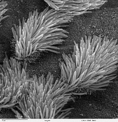
Scanning electron microscope image of lung trachea epithelium. There are both ciliated and non-ciliated cells in this epithelium. Note the difference in size between the cilia and the microvilli (on the non-ciliated cell surface). Zeiss DSM 962 SEM |
| 來源 | |
| 作者 | Charles Daghlian |
| 授權許可 (重用此檔案) |
PD |
授權條款
| 此作品已由其作者,Charles Daghlian,釋出至公有領域。此授權條款在全世界均適用。 這可能在某些國家不合法,如果是的話: Charles Daghlian授予任何人有權利使用此作品於任何用途,除受法律約束外,不受任何限制。
|
檔案歷史
點選日期/時間以檢視該時間的檔案版本。
| 日期/時間 | 縮圖 | 尺寸 | 使用者 | 備註 | |
|---|---|---|---|---|---|
| 目前 | 2006年10月7日 (六) 14:16 | | 1,024 × 1,049(375 KB) | Patho | {{Information |Description=Scanning electron microscope image of lung trachea epithelium. There are both ciliated and on-ciliated cells in this epithelium. Note the difference in size between the cilia and the microvilli(on non-ciliated cell surface) Zei |
檔案用途
下列頁面有用到此檔案:
全域檔案使用狀況
以下其他 wiki 使用了這個檔案:
- ar.wikipedia.org 的使用狀況
- ast.wikipedia.org 的使用狀況
- bs.wikipedia.org 的使用狀況
- ca.wikipedia.org 的使用狀況
- cs.wikipedia.org 的使用狀況
- da.wikipedia.org 的使用狀況
- de.wikipedia.org 的使用狀況
- de.wikibooks.org 的使用狀況
- en.wikipedia.org 的使用狀況
- es.wikipedia.org 的使用狀況
- eu.wikipedia.org 的使用狀況
- fa.wikipedia.org 的使用狀況
- fr.wikipedia.org 的使用狀況
- gl.wikipedia.org 的使用狀況
- he.wikipedia.org 的使用狀況
- he.wiktionary.org 的使用狀況
- hi.wikipedia.org 的使用狀況
- id.wikipedia.org 的使用狀況
- jv.wikipedia.org 的使用狀況
- kk.wikipedia.org 的使用狀況
- lt.wikipedia.org 的使用狀況
- lv.wikipedia.org 的使用狀況
- ms.wikipedia.org 的使用狀況
- nl.wikipedia.org 的使用狀況
- nn.wikipedia.org 的使用狀況
- no.wikipedia.org 的使用狀況
- pl.wikipedia.org 的使用狀況
- pl.wiktionary.org 的使用狀況
- pt.wikipedia.org 的使用狀況
- ru.wikipedia.org 的使用狀況
- ru.wiktionary.org 的使用狀況
檢視此檔案的更多全域使用狀況。