HL-60
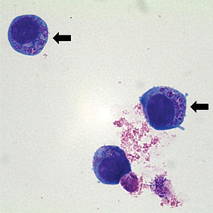
HL-60是目前已用于科学研究的人类白血病细胞系,最初是分离自一名36岁的人类女性急性早幼粒细胞白血病患者[1]。

特征
编辑HL-60细胞以悬浮培养的形式在含有营养和抗生素的培养基中持续增殖,倍增时间约为36至48小时[1]。HL-60细胞表现出嗜中性早幼粒细胞 (neutrophilic promyelocytic morphology) 形态[2],随后进行的评估则指出其出自成熟的急性骨髓性白血病。HL-60细胞通过在细胞表面表达的转铁蛋白及胰岛素受体进行增殖。如果从无血清培养基中除去转铁蛋白或胰岛素受体其中一项,HL-60细胞的增殖就会立即停止[3]。通过二甲基亚砜或维A酸的化合物,可以诱导其分化为成熟的粒细胞;而骨化三醇、佛波醇-12-十四烷酰-13-乙酸酯及颗粒白血球-巨噬细胞集落刺激因子等的化合物,则可以分别诱导HL-60细胞分化为单核细胞、巨噬细胞样或嗜酸性粒细胞表型。
科研用途
编辑HL-60细胞可以用作研究生理学、药理学和病毒学因素对髓样分化的影响。 HL-60细胞模型已经应用于研究DNA拓扑异构酶IIα和IIβ对细胞分化和凋亡的影响[4],并且特别适用于介电泳研究[5]。此外,研究人员已使用HL-60细胞来研究细胞内的钙是否在活性氧诱导的胱天蛋白酶激活中发挥到作用[6] 。HL-60细胞及衍生的分化细胞,它们的染色质和基因表达谱研究[7]是近年进行的研究。
参考资料
编辑- ^ 1.0 1.1 Gallagher, R; Collins, S; Trujillo, J; McCredie, K; Ahearn, M; Tsai, S; Metzgar, R; Aulakh, G; Ting, R; Ruscetti, F; Gallo, R. Characterization of the continuous, differentiating myeloid cell line (HL-60) from a patient with acute promyelocytic leukemia.. Blood. 1979-09, 54 (3): 713–33 [2019-12-31]. PMID 288488. (原始内容存档于2019-12-31).
- ^ Dalton WT, Jr; Ahearn, MJ; McCredie, KB; Freireich, EJ; Stass, SA; Trujillo, JM. HL-60 cell line was derived from a patient with FAB-M2 and not FAB-M3.. Blood. 1988-01, 71 (1): 242–7 [2019-12-31]. PMID 3422031. (原始内容存档于2019-12-31).
- ^ Breitman, TR; Collins, SJ; Keene, BR. Replacement of serum by insulin and transferrin supports growth and differentiation of the human promyelocytic cell line, HL-60.. Experimental cell research. 1980-04, 126 (2): 494–8 [2019-12-31]. PMID 6988226. doi:10.1016/0014-4827(80)90296-7. (原始内容存档于2019-12-31).
- ^ Sugimoto, K; Yamada, K; Egashira, M; Yazaki, Y; Hirai, H; Kikuchi, A; Oshimi, K. Temporal and spatial distribution of DNA topoisomerase II alters during proliferation, differentiation, and apoptosis in HL-60 cells.. Blood. 1998-02-15, 91 (4): 1407–17 [2019-12-31]. PMID 9454772. (原始内容存档于2019-12-31).
- ^ Ratanachoo, K; Gascoyne, PR; Ruchirawat, M. Detection of cellular responses to toxicants by dielectrophoresis.. Biochimica et biophysica acta. 2002-08-31, 1564 (2): 449–58 [2019-12-31]. PMID 12175928. doi:10.1016/s0005-2736(02)00494-7. (原始内容存档于2019-12-31).
- ^ González D., Bejarano I., Barriga C., Rodríguez A.B., Pariente J.A. Oxidative Stress-Induced Caspases are Regulated in Human Myeloid HL-60 Cells by Calcium Signal. Current Signal Transduction Therapy. 2010, 5: 181–186 [2019-12-31]. doi:10.2174/157436210791112172. (原始内容存档于2019-12-31).
- ^ Teif, VB; Mallm, JP; Sharma, T; Mark Welch, DB; Rippe, K; Eils, R; Langowski, J; Olins, AL; Olins, DE. Nucleosome repositioning during differentiation of a human myeloid leukemia cell line.. Nucleus (Austin, Tex.). 2017-03-04, 8 (2): 188–204 [2019-12-31]. PMID 28406749. doi:10.1080/19491034.2017.1295201. (原始内容存档于2019-12-31).